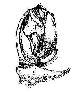
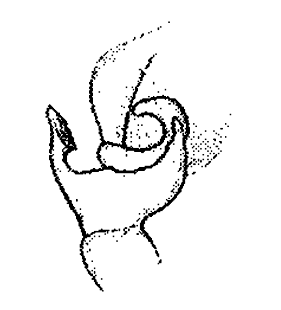
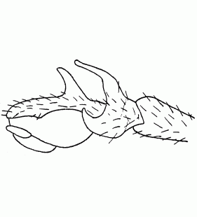
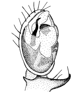
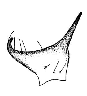
  Pedipalp Pedipalp
(Roberts 1987)
|
 Pedipalp
(Locket & Millidge 1953) |
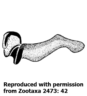
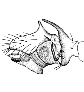
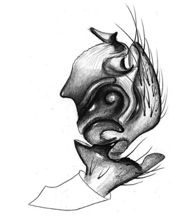 Pedipalp, retrolateral view
(Løvbrekke unpubl.) |
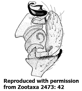
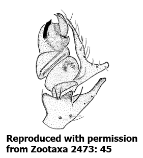
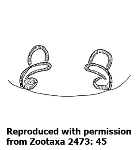
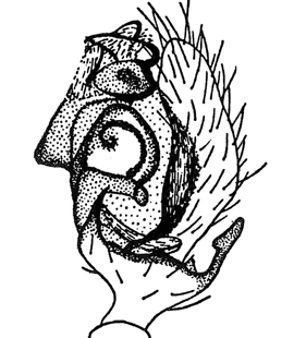
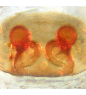
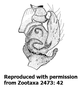 Pedipalp
(Bosmans et al. 2010) |
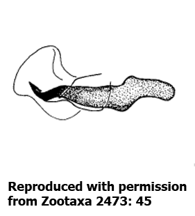
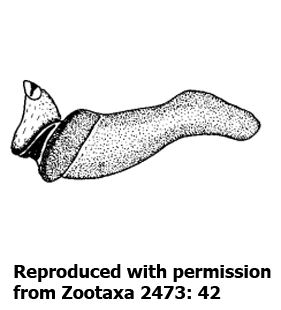 Embolic division, antero-ventral view
(Bosmans et al. 2010) |
 Palpal tibia, dorsal view
(Løvbrekke unpubl.) |
 Palpal tibia, dorsal view
(Bosmans et al. 2010) |
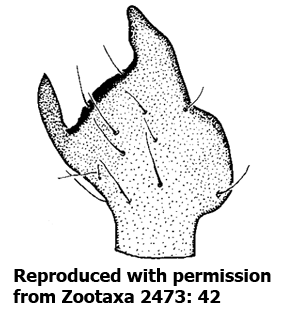
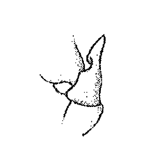
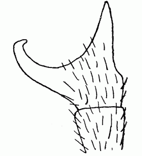
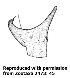
 Tibial apophysis
(Locket & Millidge 1953) |
 Tibial apophysis
(Locket & Millidge 1953) |
 Tibial apophysis
(Locket & Millidge 1953) |
 Tibial apophysis
(Locket et al. 1974) |
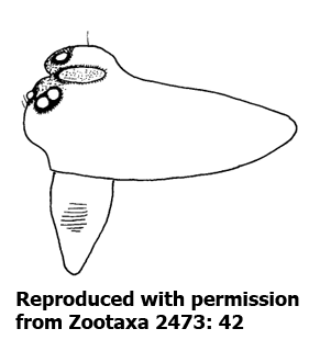
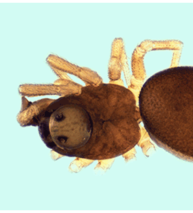
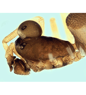
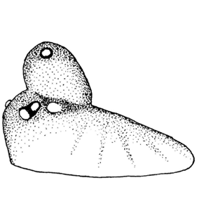
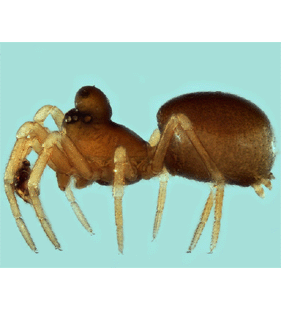
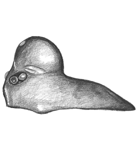
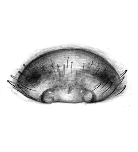
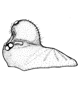 Prosoma, lateral view
(Roberts 1987) |
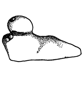 Prosoma, lateral view
(Locket & Millidge 1953) |
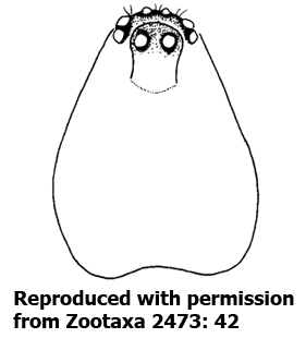
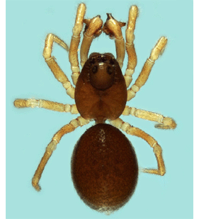
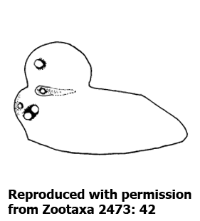
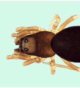
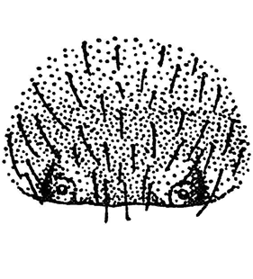
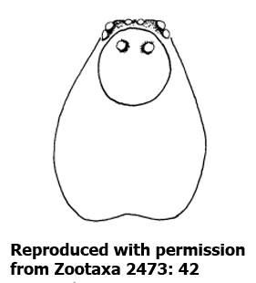 Prosoma, dorsal view
(Bosmans et al. 2010) |
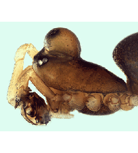
 Prosoma, lateral view
(Bosmans et al. 2010) |
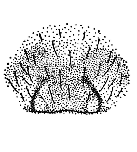
 Prosoma, dorsal view
(Oger 2020) |
 Prosoma, lateral view
(Oger 2020) |
 Prosoma, lateral view
(Løvbrekke unpubl.) |
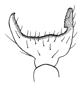
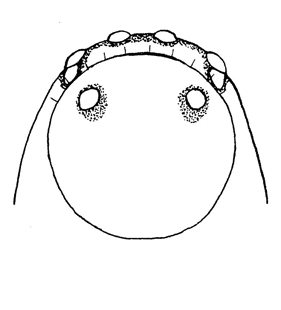
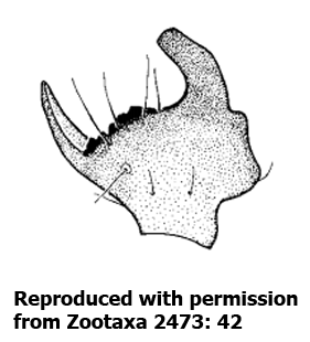
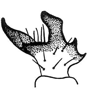
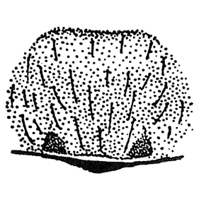
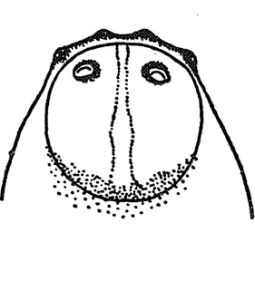 Cephalic area, dorsal view
(Locket & Millidge 1953) |
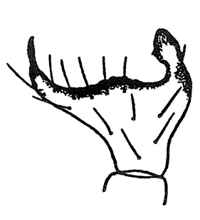
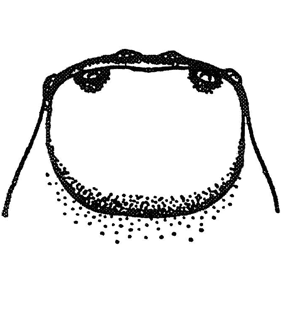 Cephalic area, dorsal view
(Locket & Millidge 1953) |
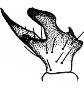
 Cephalic area, dorsal view
(Locket et al. 1974) |
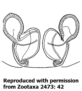
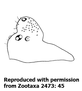
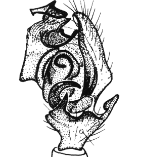
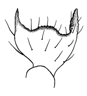
  Epigyne Epigyne
(Roberts 1987)
|
 Epigyne
(Roberts 1987) |
 Epigyne
(Roberts 1987) |
 Epigyne
(Locket & Millidge 1953) |
 Epigyne
(Locket & Millidge 1953) |
 Epigyne
(Locket & Millidge 1953) |
 Epigyne
(Løvbrekke unpubl.) |
 Epigyne, ventral view
(Bosmans et al. 2010) |
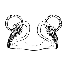
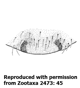
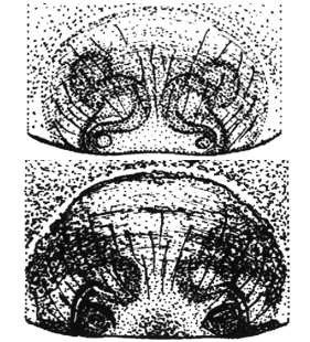
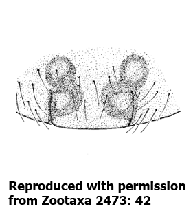
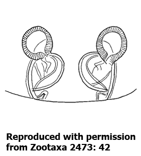
 Vulva
(Locket et al. 1974) |
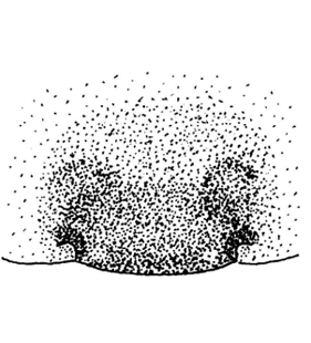
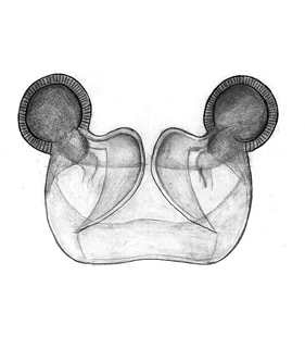 Vulva, dorsal view
(Løvbrekke unpubl.) |
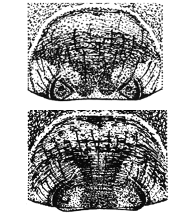
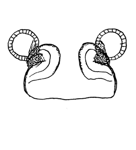
 Vulva, ventral view
(Bosmans et al. 2010) |
 Vulva, dorsal view
(Morka unpubl.) |
| |
|
|