  Pedipalp, ventral view Pedipalp, ventral view
(Roberts 1987)
|
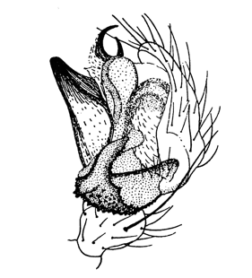  Pedipalp Pedipalp
(Locket & Millidge 1953)
|
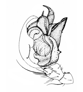  Pedipalp, retrolateral view Pedipalp, retrolateral view
(Saaristo & Wunderlich 1995)
|
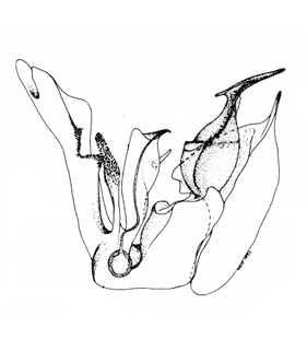  Embolic division, dorsal view Embolic division, dorsal view
(Saaristo & Wunderlich 1995)
|
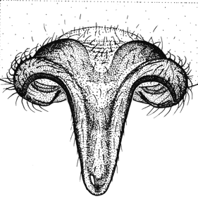  Epigyne Epigyne
(Roberts 1987)
|
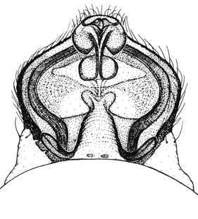  Epigyne Epigyne
(Roberts 1987)
|
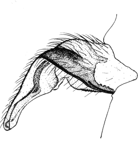  Epigyne, lateral view Epigyne, lateral view
(Roberts 1987)
|
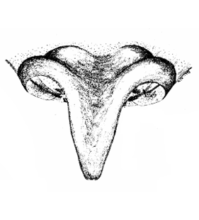  Epigyne, ventral view Epigyne, ventral view
(Saaristo & Wunderlich 1995)
|
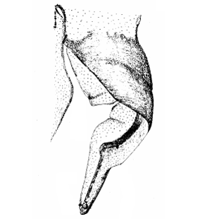  Epigyne, lateral view Epigyne, lateral view
(Saaristo & Wunderlich 1995)
|
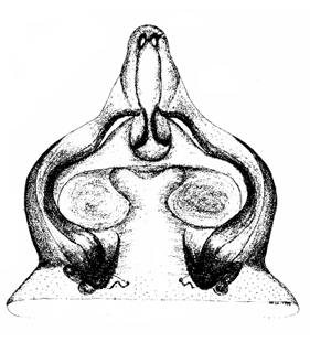  Epigyne, dorsal view Epigyne, dorsal view
(Saaristo & Wunderlich 1995)
|
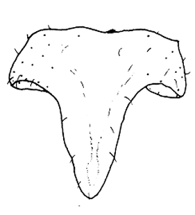  Epigyne, ventral view Epigyne, ventral view
(Růžička et al. 1991)
|
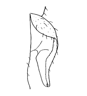  Epigyne, lateral view Epigyne, lateral view
(Růžička et al. 1991)
|
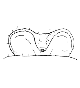  Epigyne, posterior view Epigyne, posterior view
(Růžička et al. 1991)
|
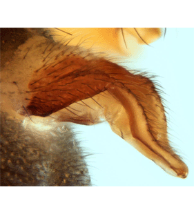  Epigyne, lateral view Epigyne, lateral view
(Janssen & Crevecoeur 2020)
|
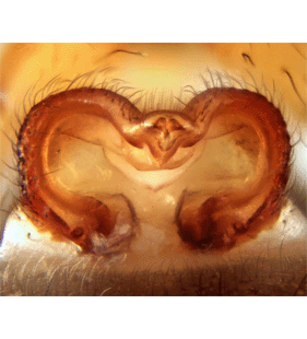  Epigyne, posterior view Epigyne, posterior view
(Janssen & Crevecoeur 2020)
|
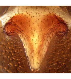  Epigyne, ventral view Epigyne, ventral view
(Janssen & Crevecoeur 2020)
|
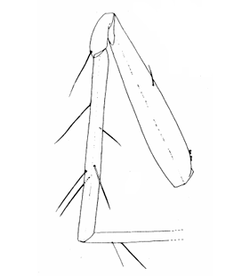  Leg I, prolateral view Leg I, prolateral view
(Saaristo & Wunderlich 1995)
|
|