|
|
In case you find an error or have a specific suggestion, please follow this link: click
|
1
|
Males 2 |
|
| - |
Females 14 |
|
2
(1)


|
Patellar apophysis missing; Prosoma as fig. female unknown Erigone jaegeri Baehr, 1984
|
|
| - |
Patellar apophysis present 3 |
|
3
(2)
|
Patellar apophysis projecting; colour light orange to yellow-brown; sternum and opisthosoma slightly blackish; eye field contrastingly black Erigone autumnalis Emerton, 1882

|
|
| - |
Patellar apophysis and colour different 4 |
|
4
(3) |
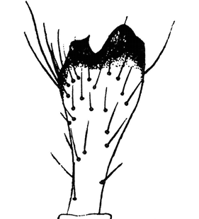
Patellar apophysis rectangularly projecting 8 |
|
| - |
Patellar apophysis projecting less than 90° from patella and directed away from cymbium 5 |
|
5
(4)
|
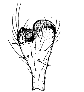
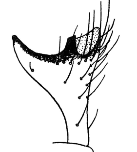
Tibia with strongly projecting apophysis Erigone svenssoni Holm, 1975
|
|
| - |
Different 6 |
|
6
(5)
|
Tibial apophysis with a distinctly bilobed part next to pointed part Erigone dentipalpis (Wider, 1834)
|
|
| - |
Different 7 |
|
7
(6)
|
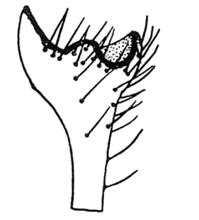
Dorsal process of tibial apophysis pointed-triangular Erigone aletris Crosby & Bishop, 1928
|
|
| -
|
Tibial apophysis with a short, stout tooth Erigone capra Simon, 1884
|
|
8
(4)
|
Patellar apophysis strikingly long, approximately rectangularly projecting, tip distinctly bent backwards Erigone psychrophila Thorell, 1871
|
|
| - |
Different 9 |
|
9
(8) |
Tibial apophysis with a median process 10 |
|
| - |
Tibial apophysis without a median process 13 |
|
10
(9) |
Median process of tibial apophysis rounded 11 |
|
| - |
Median process of tibial apophysis with one or two pointed teeth 12 |
|
11
(10)
|
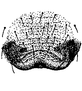
Tibial apophysis with a medially rounded, flat process Erigone atra Blackwall, 1833
|
|
| -
|
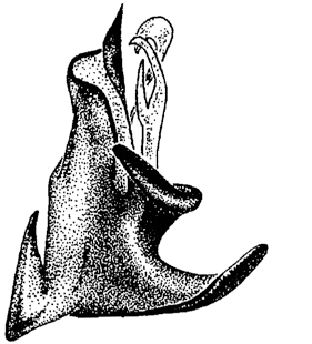
Tibial apophysis with a stronger protruding, rounded process as fig.; sides of patellar apophysis ± parallel; terminal appendix as fig. Erigone remota L. Koch, 1869
|
|
12
(10)
|
Median process of tibial apophysis with two crossing denticles Erigone longipalpis (Sundevall, 1830)
|
|
| -
|
Median process of tibial apophysis with two denticles arranged in a row (Additional remark: Holm (1956) distinguishes the north European subspecies maritima from Canadian and Sibiric populations) Erigone arctica arctica (White, 1852)
|
|
13
(9)
|
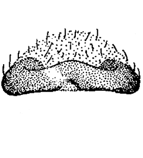
Dorsal process of tibial apophysis rounded Erigone promiscua (O. Pickard-Cambridge, 1873)
|
|
| -
|
Pedipalp as fig. Erigone tirolensis L. Koch, 1872
|
|
14
(1)
|
Epigynal plate roundish posteriorly, with median groove Erigone remota L. Koch, 1869

|
|
| - |
Different 15 |
|
15
(14) |
Posterior epigynal margin distinctly U-like indented medially or with a shallow groove 16 |
|
| - |
Posterior epigynal margin only slightly bent medially 19 |
|
16
(15)
|
Posterior epigynal margin deeply U-like indented Erigone longipalpis (Sundevall, 1830)
|
|
| - |
Posterior epigynal margin with a shallow groove 17 |
|
17
(16)
|
Lateral protruding area of epigynal plate semicircularly sclerotised Erigone capra Simon, 1884
|
|
| - |
Different 18 |
|
18
(17)
|
Epigyne lifted and in posterior view with ± triangular median part, distinctly bordered by sclerotised area anteriorly Erigone psychrophila Thorell, 1871
|
|
| -
|
Epigyne lifted and in posterior view not bordered by sclerotised area anteriorly Erigone tirolensis L. Koch, 1872
|
|
19
(15)
|
Epigynal plate laterally with two circular depressions Erigone arctica arctica (White, 1852)
|
|
| - |
Epigynal plate different, more or less unstructured 20 |
|
20
(19) |
Epigynal plate distinctly bordered laterally 21 |
|
| - |
Epigynal plate laterally different, semicircular 22 |
|
21
(20)
|
Epigynal plate almost twice as wide as long Erigone atra Blackwall, 1833
|
|
| -
|
Epigynal plate ± as long as wide Erigone dentipalpis (Wider, 1834)
|
|
22
(20) |
Posterior epigynal margin with two uniformly bent protrusions 23 |
|
| - |
Different 24 |
|
23
(22)
|
Receptacula seminis distinctly shining through epigyne; epigyne lifted and in posterior view ± quadrangular Erigone promiscua (O. Pickard-Cambridge, 1873)
|
|
| -
|
Receptacula seminis not visible through epigyne; epigyne lifted and in posterior view ± triangular Erigone aletris Crosby & Bishop, 1928
|
|
24
(22)
|
Epigyne only in posterior area with transverse folds Erigone autumnalis Emerton, 1882
|
|
| -
|
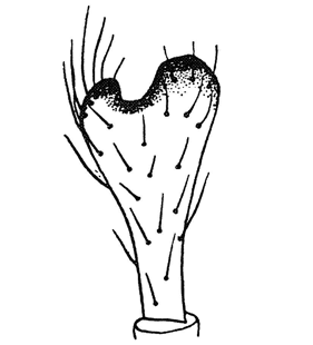
Epigyne in whole length with transverse folds Erigone svenssoni Holm, 1975
|
|
| 1. Erigone arctica palaearctica Brændegaard, 1934 |
|
| 2. Erigone arcticola Chamberlin & Ivie, 1947 |
|
| 3. Erigone cristatopalpus Simon, 1884 |
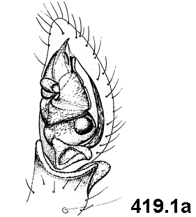
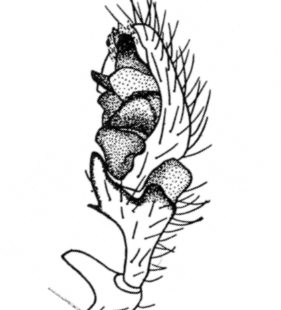
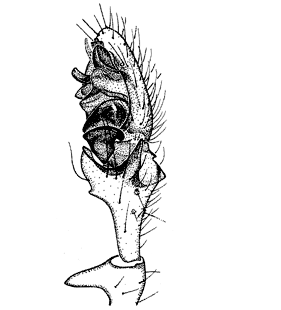
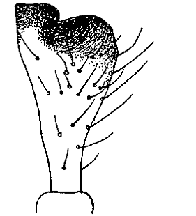
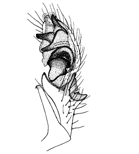
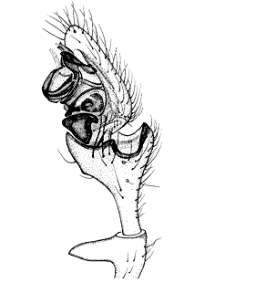
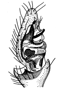
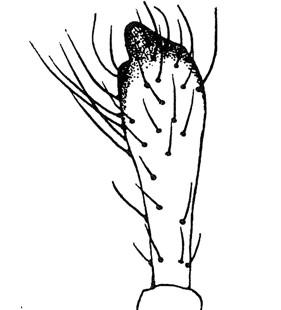
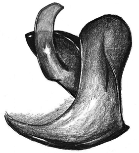
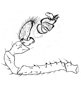
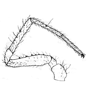
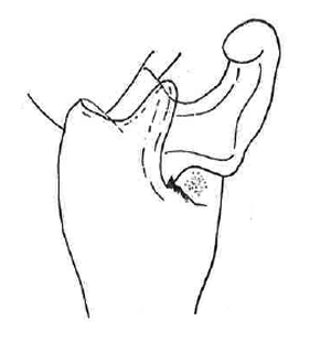
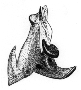
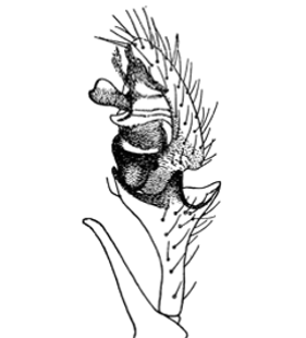
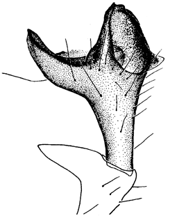
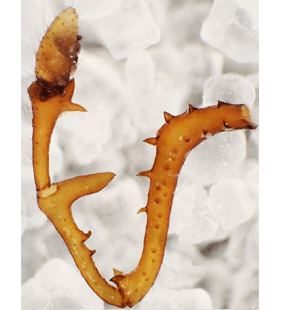
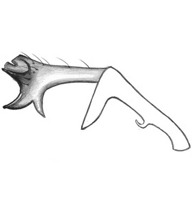
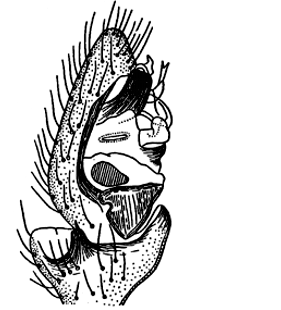
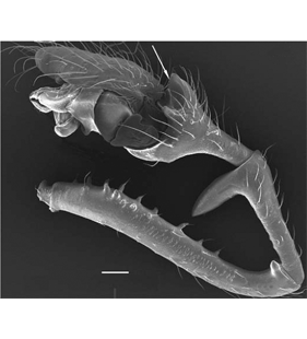
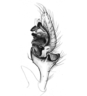
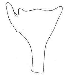
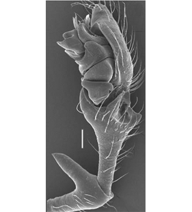
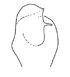
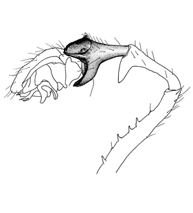 Pedipalp, retrolateral view
(Muster & Hänggi 2009) |
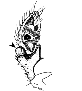
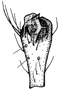
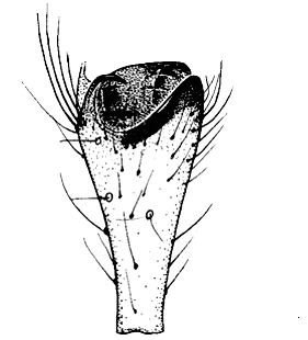
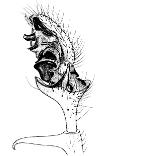
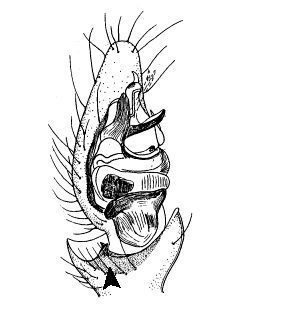
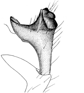
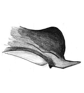
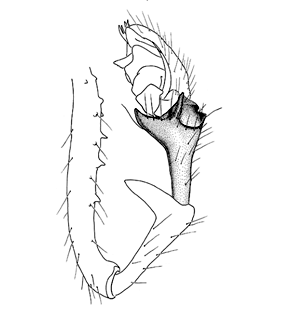
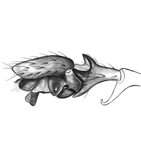
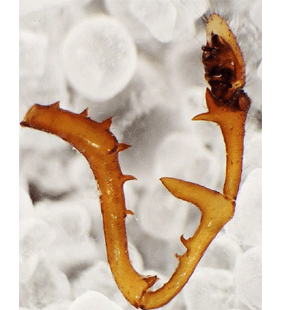
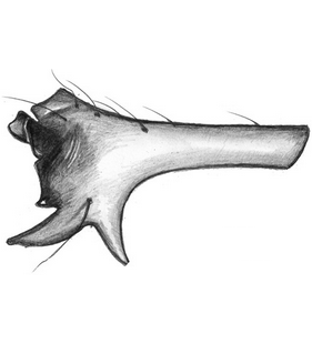
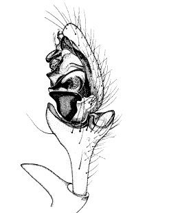
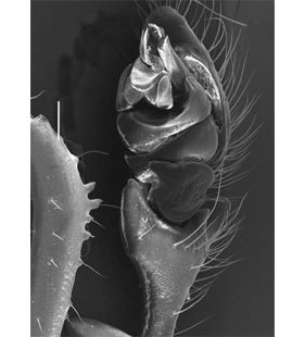
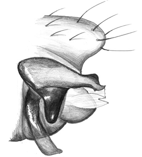
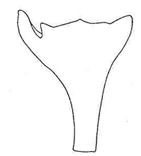
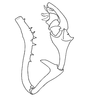
 Pedipalp, ventral view
(Muster & Hänggi 2009) |
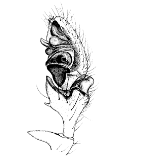
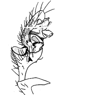
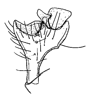
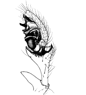
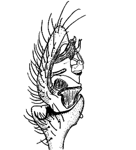
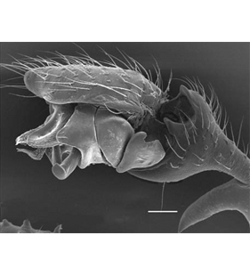
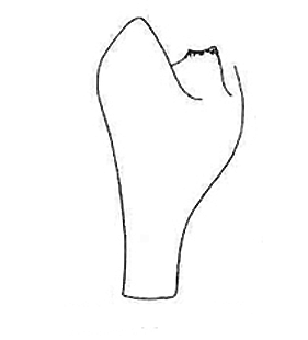
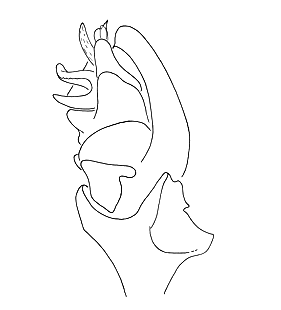
 Pedipalp, retrolateral view
(Wiśniewski & Wesołowska 2017) |
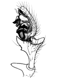
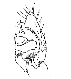
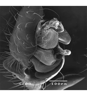
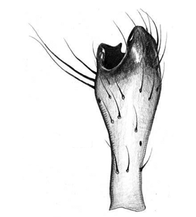
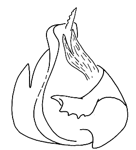
 Pedipalp, ventral view
(Wiśniewski & Wesołowska 2017) |
 Pedipalp, ventral view
(Wiśniewski & Wesołowska 2017) |
 Pedipalp, retrolateral view
(Hirna & Yanul 2023) |
 Pedipalp, ventral view
(Hirna & Yanul 2023) |
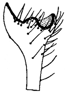
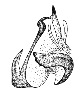
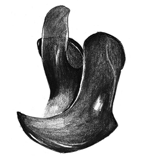
 Embolic division, prolateral-ventral view
(Muster & Hänggi 2009) |
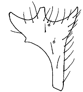
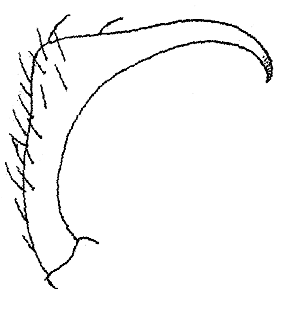
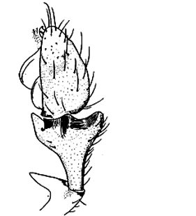
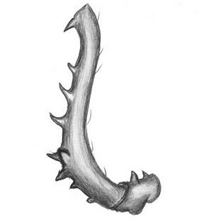
 Palpal tibia, retrolateral view
(Muster & Hänggi 2009) |
 Palpal tibia, dorsal view
(Wiśniewski & Wesołowska 2017) |
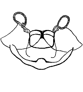
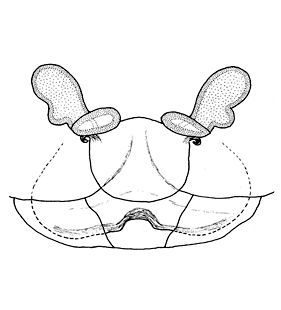
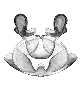
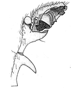
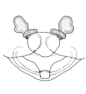
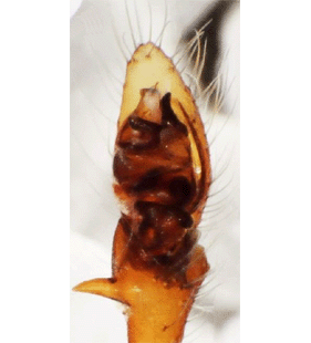
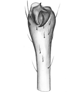
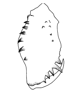 Chelicera
(Muster & Hänggi 2009) |
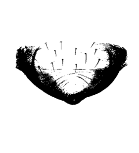
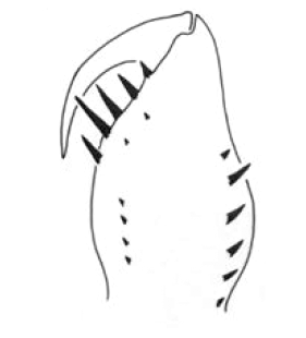 Cheliceral teeth
(Wiśniewski & Wesołowska 2017) |
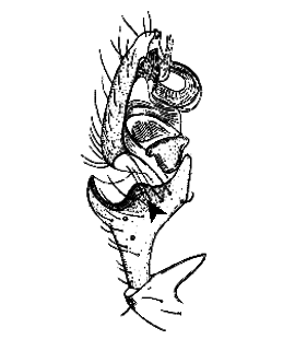
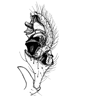
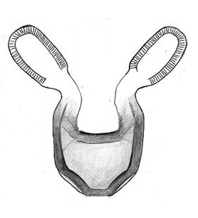
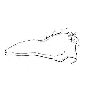
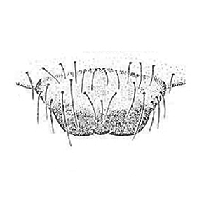
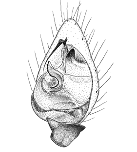
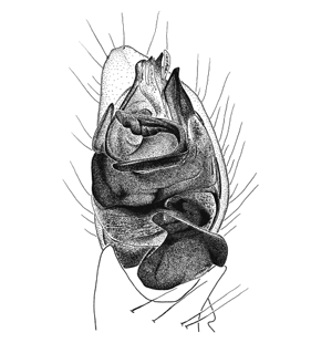
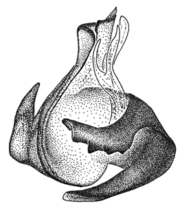
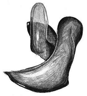
 Vulva, dorsal view
(Muster & Hänggi 2009) |
 Chelicera
(Muster & Hänggi 2009) |
|
|
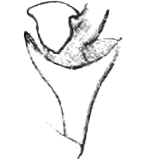
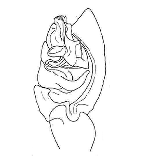
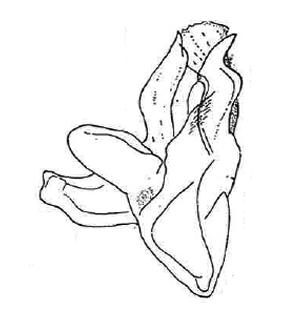
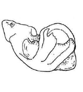
| 4. Erigone dentosa O. Pickard-Cambridge, 1894 |
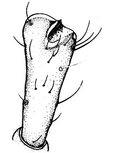
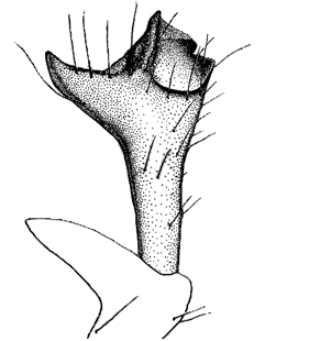
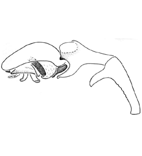
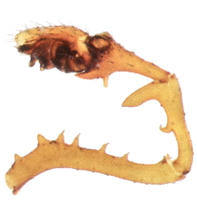
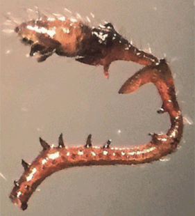
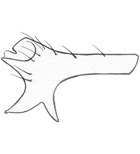
 Pedipalp, retrolateral view
(Unruh 2020) |
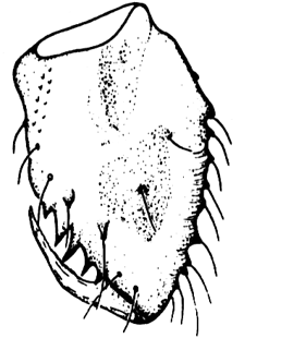
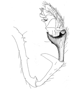
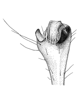
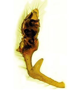
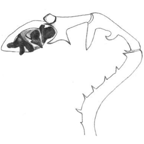
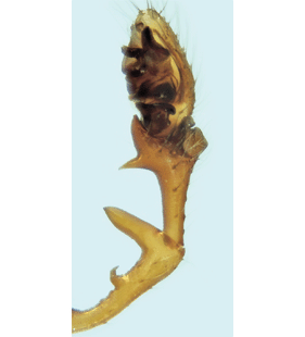
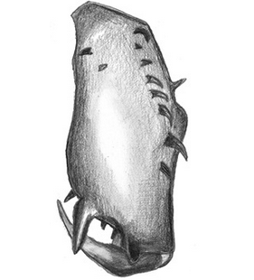
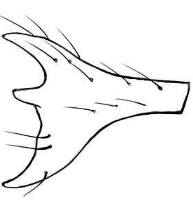
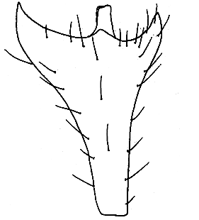
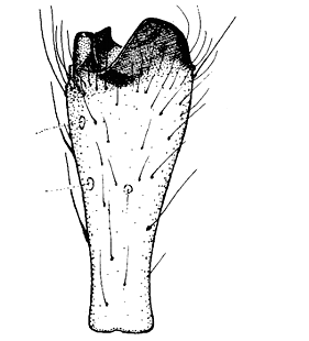
 Pedipalp with tibia, patella and femur, retrolateral view
(Arco et al. 2019) |
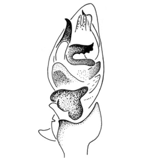
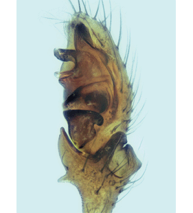
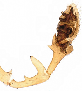
 Pedipalp with tibia, patella and femur, retrolateral view
(Crosby & Bishop 1928) |
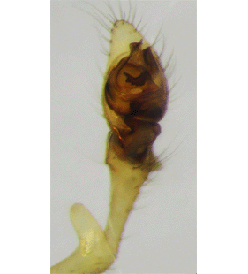
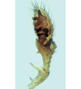
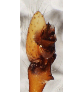
 Pedipalp with tibia, patella and femur, retrolateral view
(Kielhorn 2022) |
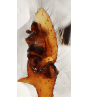
 Pedipalp with tibia, patella and femur, retrolateral view
(Berry 2022) |
 Pedipalp, retrolateral view
(Déjean & Verhoogt 2022) |
 Pedipalp with tibia and patella, retrolateral view
(Déjean & Verhoogt 2022) |
 Pedipalp with tibia and patella, retrolateral view
(Déjean & Verhoogt 2022) |
 Pedipalp with tibia and patella, retrolateral view
(Hänggi et al. 2022) |
 Pedipalp, prolateral view
(Coşar et al. 2024b) |
 Pedipalp, retrolateral view
(Coşar et al. 2024b) |
 Pedipalp with tibia, patella and femur, prolateral view
(Coşar et al. 2024b) |
 Pedipalp with tibia, patella and femur, retrolateral view
(Coşar et al. 2024b) |
 Pedipalp, ventral view
(Coşar et al. 2024b) |
 Pedipalp, ventro-prolateral view
(Coşar et al. 2024b) |
 Pedipalp with tibia, patella and femur, retrolateral view
(Martín & Adame 2024) |
 Tip of pedipalp, prolateral view
(Løvbrekke unpubl.) |
 Embolic division, anterior view
(Crosby & Bishop 1928) |
 Embolic division, mesal view
(Crosby & Bishop 1928) |
 Palpal patella, retrolateral view
(Unruh 2020) |
 Palpal tibia and patella, retrolateral view
(Løvbrekke unpubl.) |
 Palpal femur, retrolateral view
(Løvbrekke unpubl.) |
 Palpal tibia, patella and femur, retrolateral view
(Kekenbosch & Baert 2013) |
 Palpal tibia, dorsal view
(Løvbrekke unpubl.) |
 Palpal tibia, prolateral view
(Løvbrekke unpubl.) |
 Palpal tibia, retrolateral view
(Løvbrekke unpubl.) |
 Chelicerae, frontal view
(Déjean & Verhoogt 2022) |
 Chelicera, frontal view
(Løvbrekke unpubl.) |
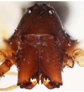 Habitus, frontal view
(Coşar et al. 2024b) |
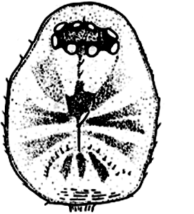
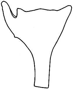
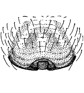
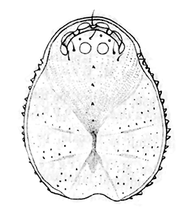
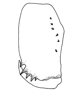
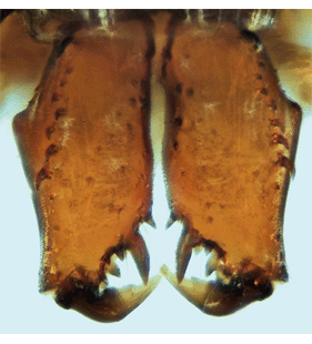
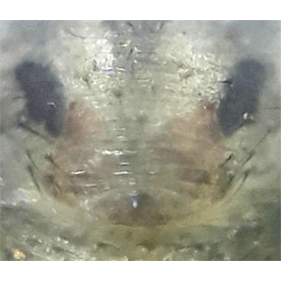
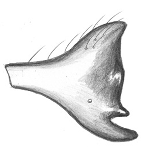
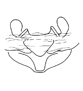
 Epigyne
(Crosby & Bishop 1928) |
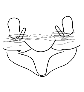
 Epigyne
(Berry 2022) |
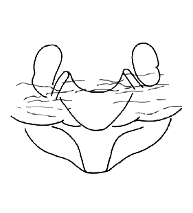
 Epigyne
(Déjean & Verhoogt 2022) |
 Epigyne, frontal view
(Løvbrekke unpubl.) |
 Vulva, dorsal view
(Berry 2022) |
 Vulva, dorsal view
(Déjean & Verhoogt 2022) |
 Vulva, dorsal view
(Hänggi et al. 2022) |
 Vulva, dorsal view
(Unruh 2020) |
 Vulva, ventral view
(Løvbrekke unpubl.) |
 Vulva, dorsal view
(Løvbrekke unpubl.) |
| |
|
|
|
| 5. Erigone dumitrescuae Georgescu, 1969 |
|
| 6. Erigone hypoarctica Eskov, 1989 |
|
| 7. Erigone jugorum Simon, 1884 |
|
| 8. Erigone longipalpis pirini Deltshev, 1983 |
|
| 9. Erigone maritima Kulczyński, 1902 |
 Pedipalp
(Tyschchenko 1971) |
 Pedipalp
(Roberts 1987) |
 Pedipalp
(Wiehle 1960a) |
 Pedipalp, retrolateral view
(Marusik et al. 2006b) |
 Pedipalp, retrolateral view
(Marusik et al. 2006b) |
 Pedipalp, ventral view
(Marusik et al. 2006b) |
 Pedipalp in detail
(Marusik et al. 2006b) |
 Pedipalp, retrolateral view
(Løvbrekke unpubl.) |
 Apical part of bulbus, dorsal view
(Løvbrekke unpubl.) |
 Apical part of bulbus, dorsal view
(Løvbrekke unpubl.) |
 Tip of pedipalp, prolateral view
(Løvbrekke unpubl.) |
 Embolus, dorsal view
(Løvbrekke unpubl.) |
 Embolic division, prolateral view
(Løvbrekke unpubl.) |
 Lateral apophysis
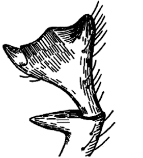
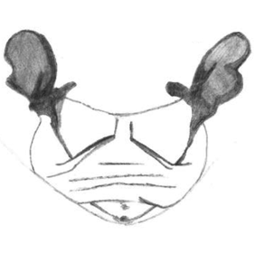
(Wiehle 1960a) |
 Patellar and tibial apophysis
(Wiehle 1960a) |
 Palpal tibia and patella, retrolateral view
(Løvbrekke unpubl.) |
 Palpal tibia
(Holm 1956) |
 Palpal tibia
(Holm 1956) |
 Palpal tibia
(Holm 1956) |
 Palpal tibia, dorsal view
(Løvbrekke unpubl.) |
 Palpal tibia, prolateral view
(Løvbrekke unpubl.) |
 Palpal tibia, retrolateral view
(Løvbrekke unpubl.) |
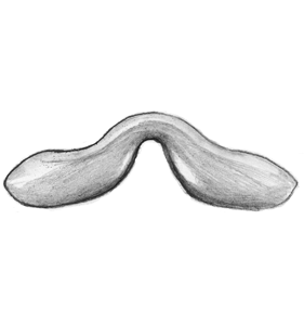
 Tibial apophysis
(Locket et al. 1974) |
 Tibial apophysis
(Roberts 1987) |
 Tibial apophysis
(Locket & Millidge 1953) |
 Tibial apophysis
(Locket & Millidge 1953) |
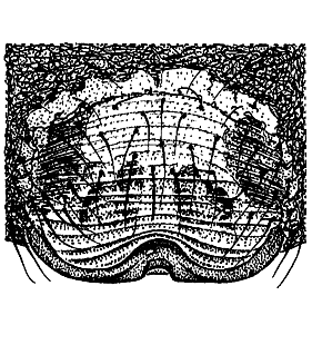
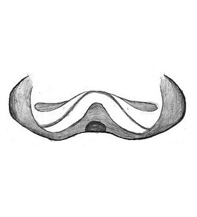
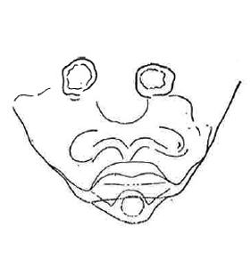
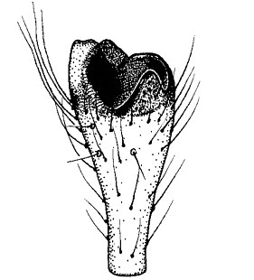
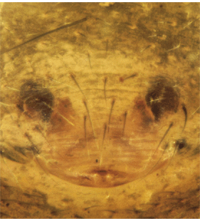
 Epigyne
(Locket & Millidge 1953) |
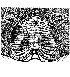
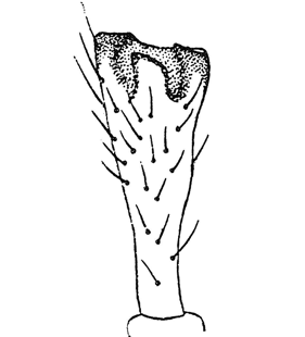
 Epigyne
(Locket & Millidge 1953) |
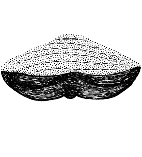
 Epigyne
(Roberts 1987) |
 Epigyne, lifted
(Roberts 1987) |
 Epigyne
(Tyschchenko 1971) |
 Epigyne
(Wiehle 1960a) |
 Epigyne, ventral view
(Løvbrekke unpubl.) |
 Epigyne, ventral view
(Løvbrekke unpubl.) |
 Epigyne
(Løvbrekke unpubl.) |
 Epigyne, frontal view
(Løvbrekke unpubl.) |
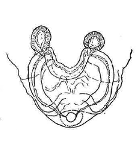
 Vulva
(Locket et al. 1974) |
 Vulva
(Holm 1956) |
 Vulva
(Wiehle 1960a) |
 Vulva, dorsal view
(Løvbrekke unpubl.) |
 Vulva, dorsal view
(Løvbrekke unpubl.) |
|
|
| 10. Erigone nigrimana Thorell, 1875 |
 -- Caption missing --
(WSC 2025) |
|
|
|
| 11. Erigone remota dentigera Simon, 1926 |
|
| 12. Erigone tenuimana Simon, 1884 |
|
| 13. Erigone welchi Jackson, 1911 |
|
| 14. Erigone whymperi O. Pickard-Cambridge, 1877 |
 Pedipalp, ventral view
(Marusik et al. 2006b) |
 Pedipalp, retrolateral view
(Marusik et al. 2006b) |
 Pedipalp, retrolateral view
(Marusik et al. 2006b) |
 Pedipalp, lateral view
(Tanasevitch & Koponen 2007) |
 Pedipalp
(Tanasevitch & Koponen 2007) |
 Embolic division
(Tanasevitch & Koponen 2007) |
 Embolic division
(Tanasevitch & Koponen 2007) |
 Tooth of embolic division
(Tanasevitch & Koponen 2007) |
 Tooth of embolic division
(Tanasevitch & Koponen 2007) |
 Tooth of embolic division
(Tanasevitch & Koponen 2007) |
 Tooth of embolic division
(Tanasevitch & Koponen 2007) |
 Palpal femur
(Tanasevitch & Koponen 2007) |
 Palpal tibia, lateral view
(Tanasevitch & Koponen 2007) |
 Palpal tibia, lateral view
(Tanasevitch & Koponen 2007) |
 Palpal tibia, lateral view
(Tanasevitch & Koponen 2007) |
 Palpal tibia, lateral view
(Tanasevitch & Koponen 2007) |
 Palpal tibia
(Tanasevitch & Koponen 2007) |
 Epigyne, dorsal view
(Tanasevitch & Koponen 2007) |
 Epigyne, dorsal view
(Tanasevitch & Koponen 2007) |
 Epigyne, dorsal view
(Tanasevitch & Koponen 2007) |
 Epigyne, dorsal view
(Tanasevitch & Koponen 2007) |
 Vulva
(Tanasevitch & Koponen 2007) |
 Vulva
(Tanasevitch & Koponen 2007) |
|
|