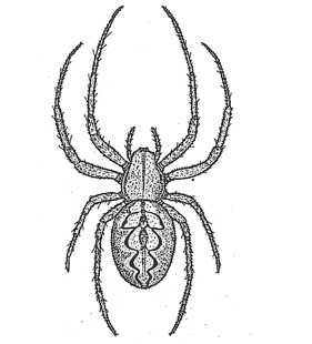
| 1. Neoscona byzanthina (Pavesi, 1876) |
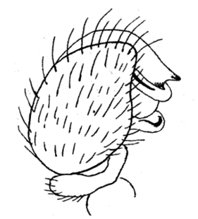
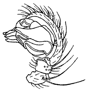
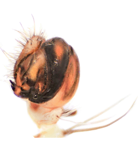
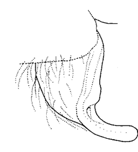
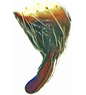
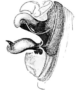
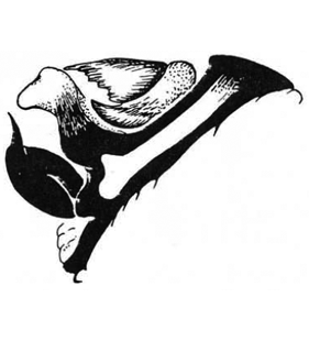
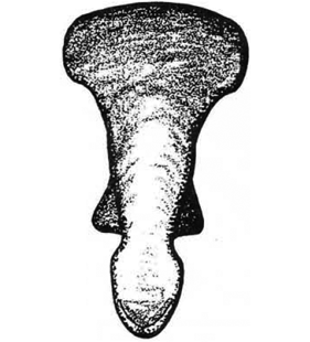
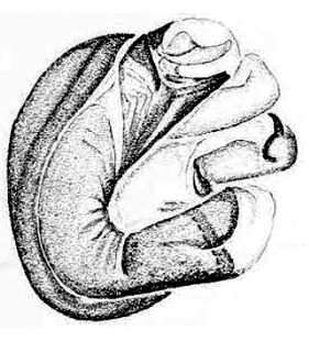
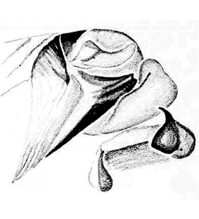
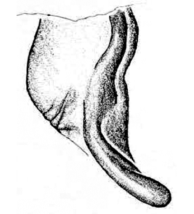
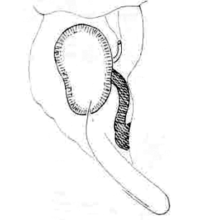
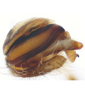
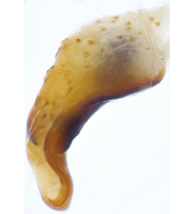
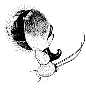
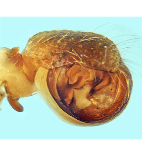
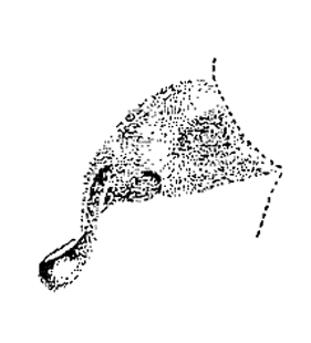
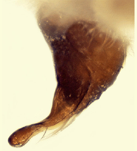
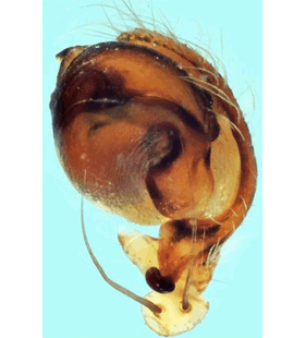
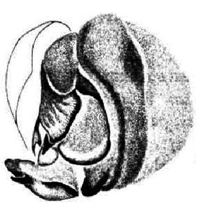
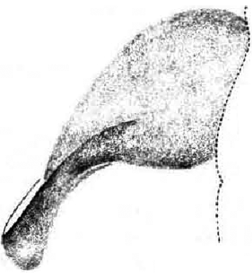
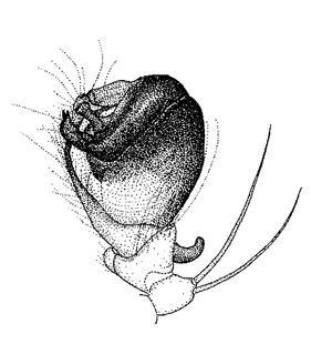  Pedipalp, retrolateral view Pedipalp, retrolateral view
(Ledoux 2008a)
|
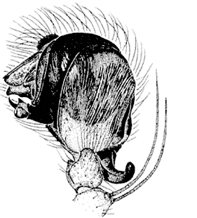
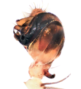
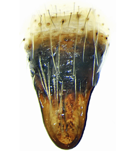
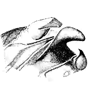
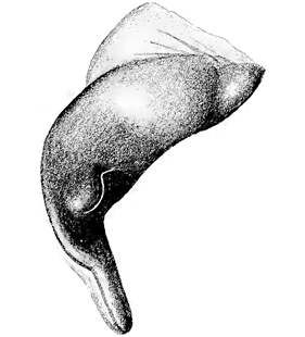
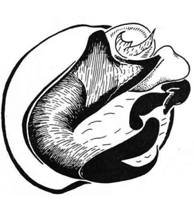
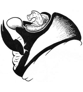
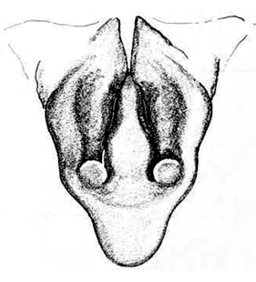
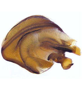
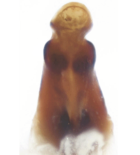
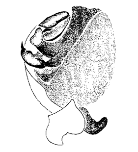
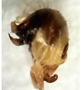
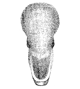
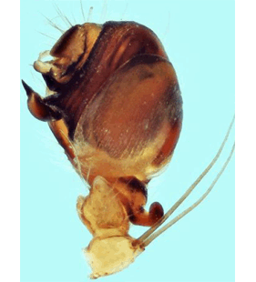
 Pedipalp, apical view
(Geci & Naumova 2021a) |
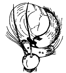
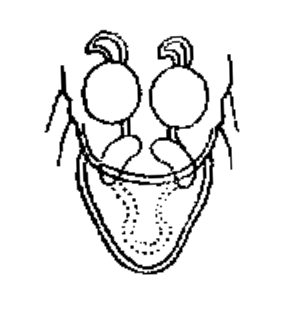
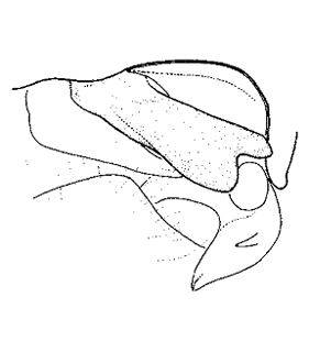
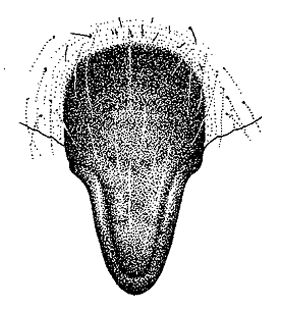
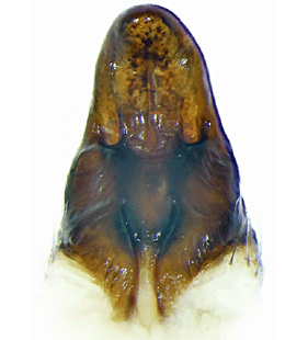
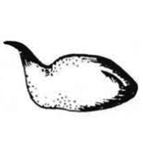
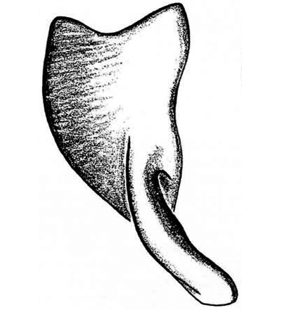
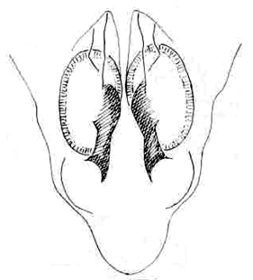
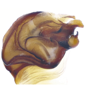
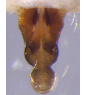
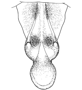
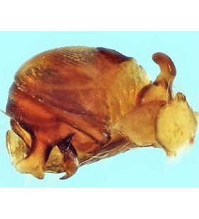
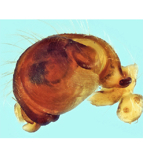
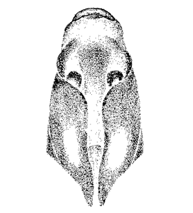
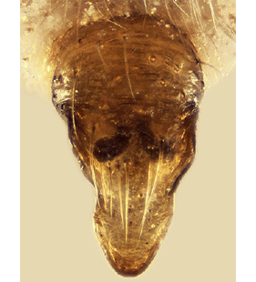
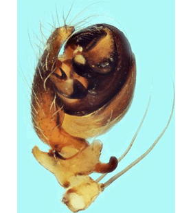
 Pedipalp, ventral view
(Geci & Naumova 2021a) |
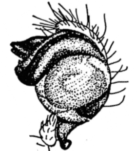
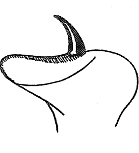
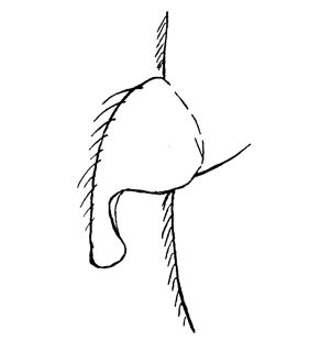
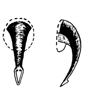
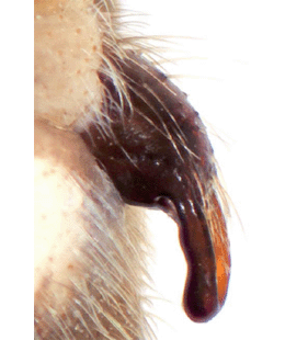
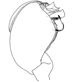
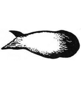
 Tip of bulbus
(Ledoux 2008a) |
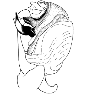
 Tip of bulbus
(Ledoux 2008a) |
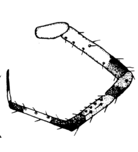
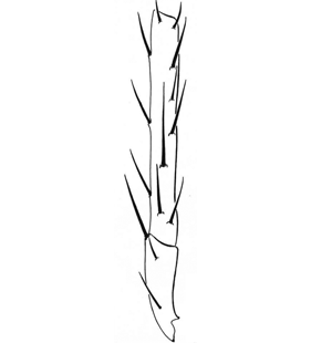
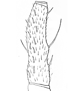
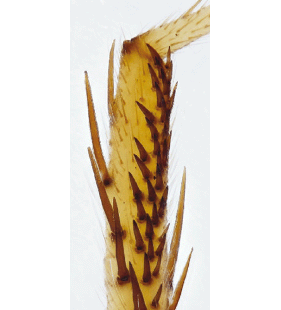
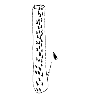
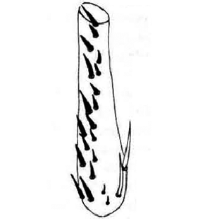
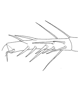 Tibia II, prolateral view
(Ledoux 2008a) |
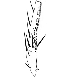
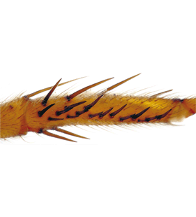
 Tibia II, prolateral view
(Geci & Naumova 2021a) |
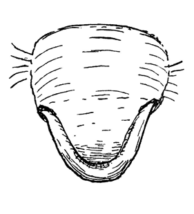
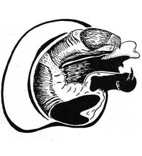
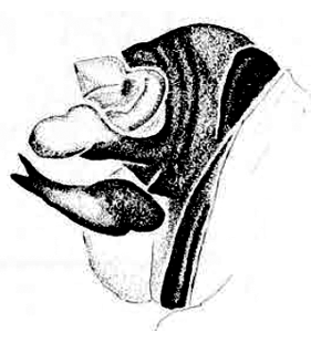
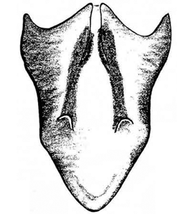
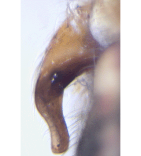
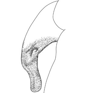
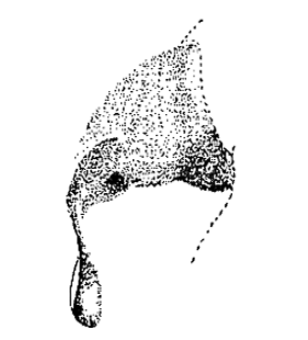
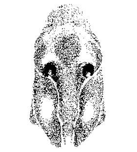
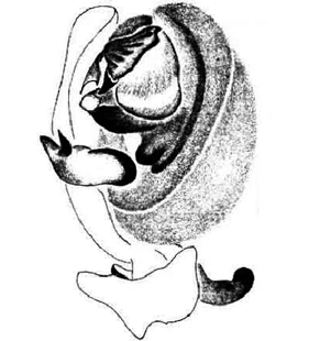
  Epigyne, lateral view Epigyne, lateral view
(Ledoux 2008a)
|
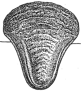
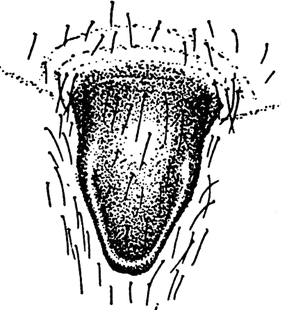
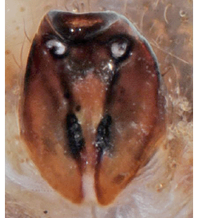
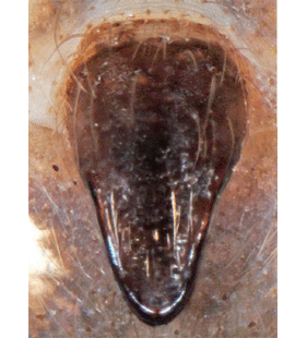
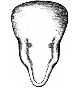
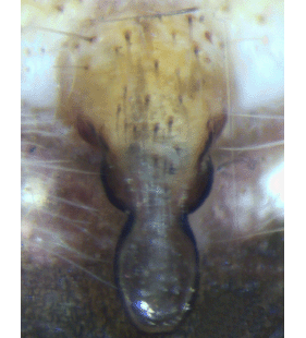
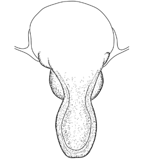
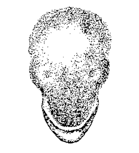
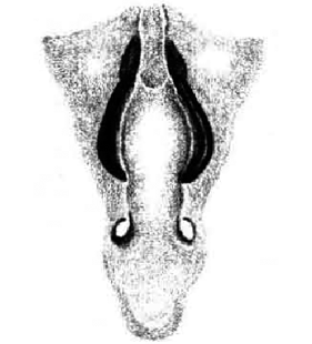
 Epigyne, ventral view
(Ledoux 2008a) |
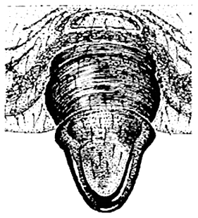
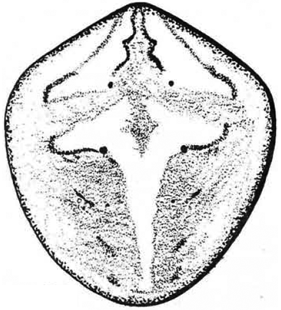
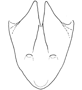
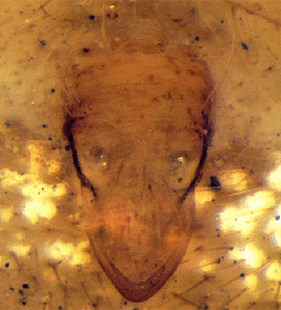
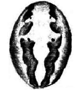
 Epigyne, posterior view
(Mora-Rubio et al. 2019) |
 Epigyne, lateral view
(Geci & Naumova 2021a) |
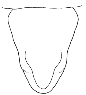
 Epigyne, posterior view
(Geci & Naumova 2021a) |
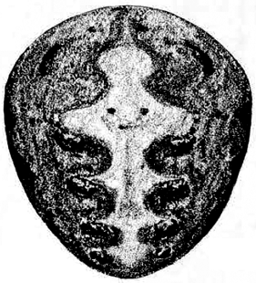
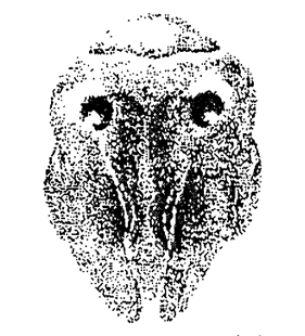
 Epigyne, ventral view
(Geci & Naumova 2021a) |
 Epigyne, lateral view
(Pintilioaie & Urák 2022a) |
 Epigyne, posterior view
(Pintilioaie & Urák 2022a) |
 Epigyne, ventral view
(Pintilioaie & Urák 2022a) |
|
|
|
| 2. Neoscona crucifera (Lucas, 1838) |
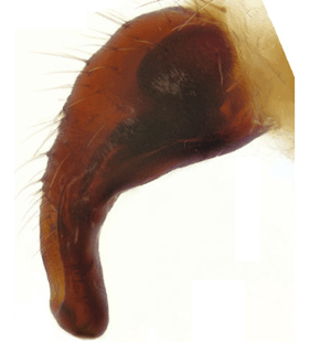
 Pedipalp, ventral view
(Berman & Levi 1971) |
 Pedipalp, apical view
(Berman & Levi 1971) |
 Tip of pedipalp, lateral view
(Berman & Levi 1971) |
 Tip of pedipalp, ventral view
(Berman & Levi 1971) |
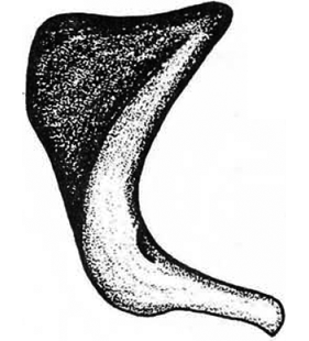
 Conductor, lateral view
(Grasshoff 1986) |
 Terminal apophysis
(Grasshoff 1986) |
 Terminal apophysis
(Grasshoff 1986) |
 Tibia II, ventro-prolateral view
(Berman & Levi 1971) |
 Tibia I
(Grasshoff 1986) |
 Tibia II
(Grasshoff 1986) |
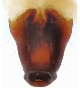
 Epigyne, lateral view
(Berman & Levi 1971) |
 Epigyne, posterior view
(Berman & Levi 1971) |
 Epigyne, ventral view
(Berman & Levi 1971) |
 Epigyne, lateral view
(Grasshoff 1986) |
 Epigyne, posterior view
(Grasshoff 1986) |
 Epigyne, ventral view
(Grasshoff 1986) |
 Epigyne, ventral view
(Grasshoff 1986) |
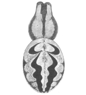
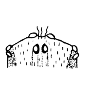
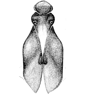
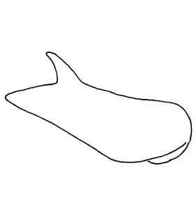
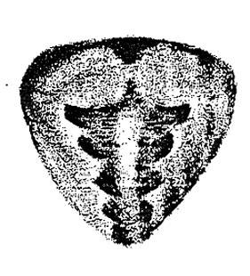
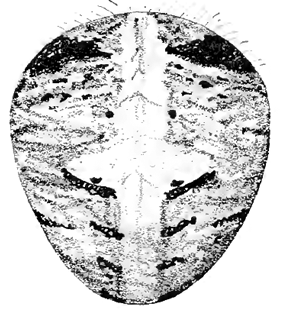 Opisthosoma, dorsal view
(Berman & Levi 1971) |
 Opisthosoma, dorsal view
(Grasshoff 1986) |
 Opisthosoma, dorsal view
(Grasshoff 1986) |
|
|
| 3. Neoscona nautica (L. Koch, 1875) |
 Pedipalp, apical view
(Berman & Levi 1971) |
 Pedipalp, ventral view
(Berman & Levi 1971) |
 Pedipalp, ventral view
(Grasshoff 1986) |
 Pedipalp, dorsal view
(Tanikawa 1998) |
 Pedipalp, ventral view
(Tanikawa 1998) |
 Tip of pedipalp, ventral view
(Berman & Levi 1971) |
 Tip of pedipalp, lateral view
(Berman & Levi 1971) |
 Tip of pedipalp, lateral view
(Grasshoff 1986) |
 Conductor, lateral view
(Grasshoff 1986) |
 Conductor, lateral view
(Tanikawa 1998) |
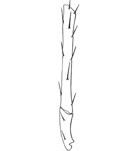
 Tibia II, ventro-prolateral view
(Berman & Levi 1971) |
 Tibia I
(Grasshoff 1986) |
 Tibia II
(Grasshoff 1986) |
 Tibia II, prolateral view
(Tanikawa 1998) |
 Epigyne, lateral view
(Berman & Levi 1971) |
 Epigyne, posterior view
(Berman & Levi 1971) |
 Epigyne, lateral view
(Grasshoff 1986) |
 Epigyne, posterior view
(Grasshoff 1986) |
 Epigyne, ventral view
(Grasshoff 1986) |
 Epigyne, posterior view
(Tanikawa 1998) |
 Epigyne, lateral view
(Tanikawa 1998) |
 Epigyne, ventral view
(Tanikawa 1998) |
 Vulva, ventral view
(Berman & Levi 1971) |
 Vulva, lateral view
(Berman & Levi 1971) |
 Vulva, posterior view
(Berman & Levi 1971) |
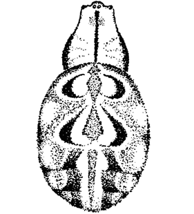
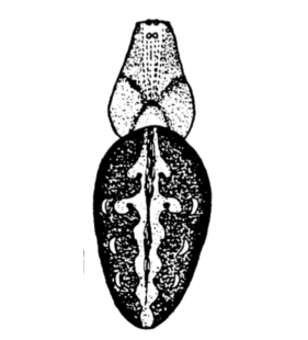
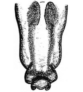
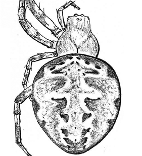
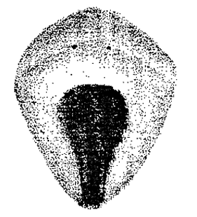
 Opisthosoma, dorsal view
(Berman & Levi 1971) |
 Opisthosoma, ventral view
(Berman & Levi 1971) |
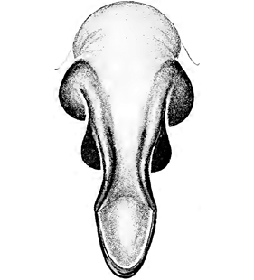
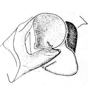
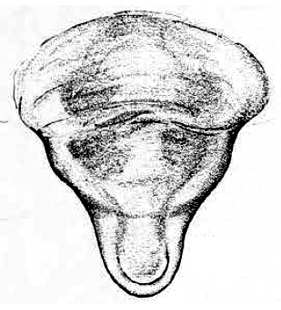
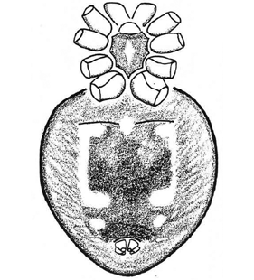
 Habitus, dorsal view
(Grasshoff 1986) |
 Habitus, ventral view
(Grasshoff 1986) |
|
|
| 4. Neoscona spasskyi (Brignoli, 1983) |
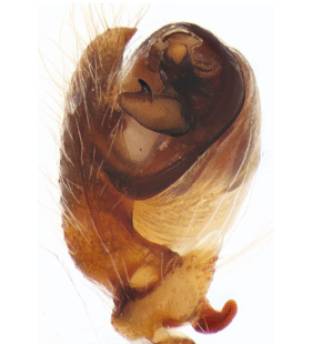 Pedipalp, prolateral view
(Zamani et al. 2020a) |
 Bulbus, anterior view
(Zamani et al. 2020a) |
 Bulbus, ventral view
(Zamani et al. 2020a) |
 Bulbus, ventral view
(Zamani et al. 2020a) |
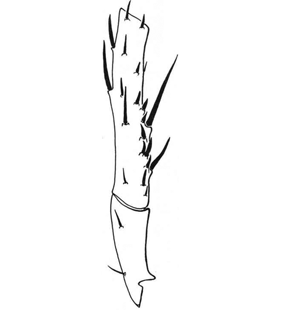
 Tibia II, ventral view
(Zamani et al. 2020a) |
 Epigyne, lateral view
(Zamani et al. 2020a) |
 Epigyne, posterior view
(Zamani et al. 2020a) |
 Epigyne, ventral view
(Zamani et al. 2020a) |
 Epigyne, dorsal view
(Seropian et al. 2023a) |
 Epigyne, dorsal view
(Seropian et al. 2023a) |
 Epigyne, lateral view
(Seropian et al. 2023a) |
 Epigyne, lateral view
(Seropian et al. 2023a) |
 Epigyne, ventral view
(Seropian et al. 2023a) |
 Epigyne, ventral view
(Seropian et al. 2023a) |
|
|
| 5. Neoscona subfusca (C. L. Koch, 1837) |
 Pedipalp, ventral view
(Pesarini unpubl.) |
 Pedipalp, prolateral view
(Levy 1998a) |
 Pedipalp, ventral view
(Levy 1998a) |
 Pedipalp, prolateral view
(Bosmans & Hervé 2021) |
 Pedipalp, retrolateral view
(Bosmans & Hervé 2021) |
 Pedipalp, ventro-retrolateral view
(Bosmans & Hervé 2021) |
 Pedipalp, prolateral view
(Stanković 2023) |
 Tibia II
(Levy 1998a) |
 Epigyne, lateral view
(Levy 1998a) |
 Epigyne, lateral view
(Levy 1998a) |
 Epigyne, ventral view
(Levy 1998a) |
 Epigyne, posterior view
(Levy 1998a) |
 Epigyne
(Bosmans & Hervé 2021) |
 Epigyne, lateral view
(Pintilioaie & Urák 2022a) |
 Epigyne, posterior view
(Pintilioaie & Urák 2022a) |
 Epigyne, ventral view
(Pintilioaie & Urák 2022a) |
 Epigyne, variation, ventral view
(Levy 1998a) |
 Epigyne, variation, posterior view
(Levy 1998a) |
 Epigyne, variation, posterior view
(Levy 1998a) |
 Opisthosoma, dorsal view
(Levy 1998a) |
 Opisthosoma, dorsal view
(Levy 1998a) |
| |
|
|
|
| 6. Neoscona tedgenica (Bakhvalov, 1978) |
|
| 7. Neoscona theisi (Walckenaer, 1841) |
 Pedipalp, prolateral view
(Bosmans et al. 2019c) |
 Pedipalp, retrolateral view
(Bosmans et al. 2019c) |
 Pedipalp, ventral view
(Bosmans et al. 2019c) |
 Pedipalp, ventro-prolateral view
(Bosmans et al. 2019c) |
 Pedipalp, apical view
(Levy 1998a) |
 Pedipalp, mesal view
(Levy 1998a) |
 Pedipalp, prolateral view
(Levy 1998a) |
 Pedipalp, ventral view
(Levy 1998a) |
 Pedipalp, prolateral view
(Zamani et al. 2020a) |
 Tibia II
(Levy 1998a) |
 Tibia II, ventral view
(Zamani et al. 2020a) |
 Opisthosoma, dorsal view
(Levy 1998a) |
 Epigyne, lateral view
(Levy 1998a) |
 Epigyne, posterior view
(Levy 1998a) |
 Epigyne, ventral view
(Levy 1998a) |
 Epigyne, lateral view
(Zamani et al. 2020a) |
 Epigyne, posterior view
(Zamani et al. 2020a) |
 Epigyne, ventral view
(Zamani et al. 2020a) |
 Sternum
(Zamani et al. 2020a) |
 Opisthosoma, dorsal view
(Levy 1998a) |
 Opisthosoma, ventral view
(Zamani et al. 2020a) |
| |
|
|
|