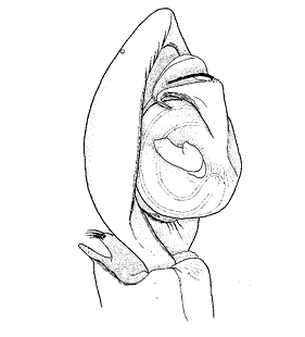
  Pedipalp, retrolateral view Pedipalp, retrolateral view
(Thaler & Knoflach 1998a)
|
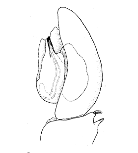
 Pedipalp, ventral view
(Thaler & Knoflach 1998a) |
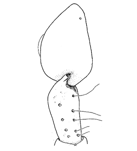
 Pedipalp, mesal view
(Thaler & Knoflach 1998a) |
 Pedipalp, dorsal view
(Thaler & Knoflach 1998a) |
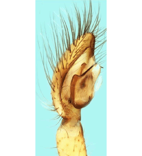 Pedipalp, prolateral view
(Bosmans et al. 2019c) |
 Pedipalp, retrolateral view
(Bosmans et al. 2019c) |
 Pedipalp, ventral view
(Bosmans et al. 2019c) |
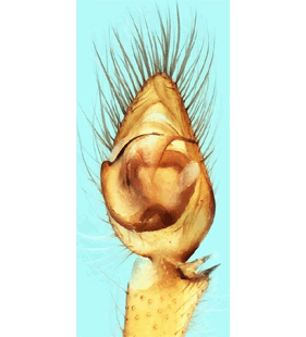
 Embolus and conductor
(Thaler & Knoflach 1998a) |
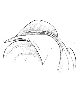
 Palpal tibia, dorsal view
(Thaler & Knoflach 1998a) |
 Palpal tibia, dorsal view
(Bosmans et al. 2019c) |
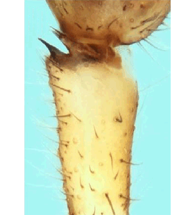
 Metatarsus I
(Thaler & Knoflach 1998a) |
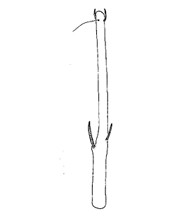 Metatarsus I
(Thaler & Knoflach 1998a) |
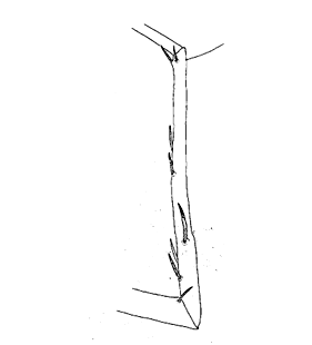
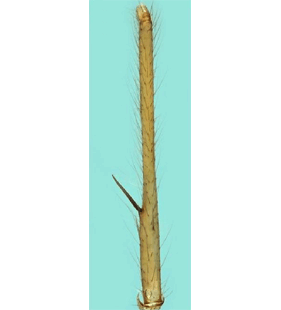 Metatarsus I
(Bosmans et al. 2019c) |
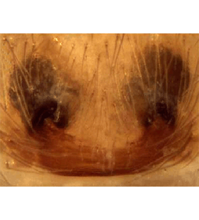
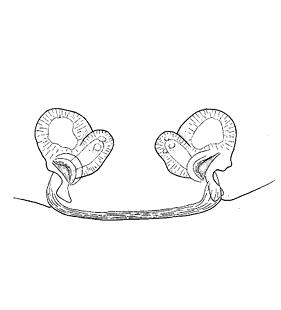  Epigyne/vulva, ventral view Epigyne/vulva, ventral view
(Thaler & Knoflach 1998a)
|
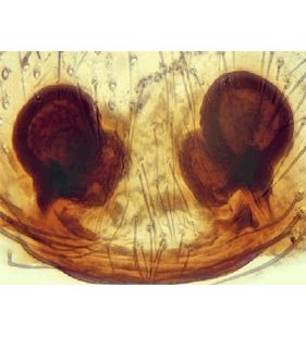
 Epigyne/vulva, dorsal view
(Thaler & Knoflach 1998a) |
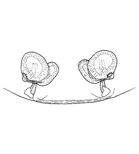
 Epigyne
(Bosmans et al. 2019c) |
 Vulva
(Bosmans et al. 2019c) |
|
